Your Trusted Veterinarian in Golden, CO
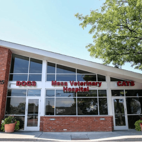
Welcome to Mesa Veterinary Hospital in Golden, CO
Celebrating 50 years in Golden, CO’s Applewood neighborhood, Mesa Veterinary Hospital has built lasting relationships with generations of clients and their beloved pets. We invite you and your pet to become part of the Mesa family, and we look forward to helping you both enjoy many happy years together.
We are AAHA Accredited

About Us
About Mesa Veterinary Hospital
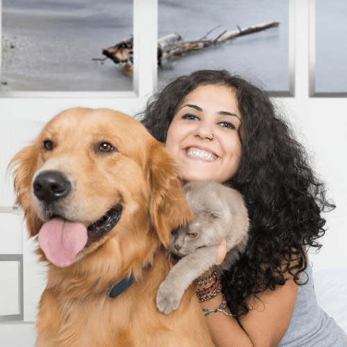
At Mesa Veterinary Hospital, we value the special relationships that we build with our clients and their pets. We know what a privilege it is to be welcomed into your family, and we strive to nurture that bond each time you need us. The families and patients we are able to serve are extraordinary, so we want to ensure that the experience you have here with us is extraordinary, too!
We are a nine-veterinarian full-service small animal and exotic veterinary hospital in West Denver, Colorado. At Mesa Veterinary Hospital, we celebrate and nurture the special role your pet has in your life. Alongside our experienced clinical and administrative team, we strive to provide compassionate, progressive, and exceptional veterinary care.
At Mesa, we value the special relationships that we build with our clients and their pets.
Complete our various patient forms right from our website and save time visiting our practice.
We offer a range of comprehensive services to ensure your pets receive the best care.
Our veterinary team is here for you and your pet. Reach out anytime with questions.

Thank you for your kind words
Full-Service Veterinary Care
Complete Veterinary Services in Golden, CO
We provide a variety of comprehensive services to fit your needs. See a complete list of our services and additional information below.

Pet Surgery
We specialize in providing exceptional quality veterinary surgery services for your beloved pets.

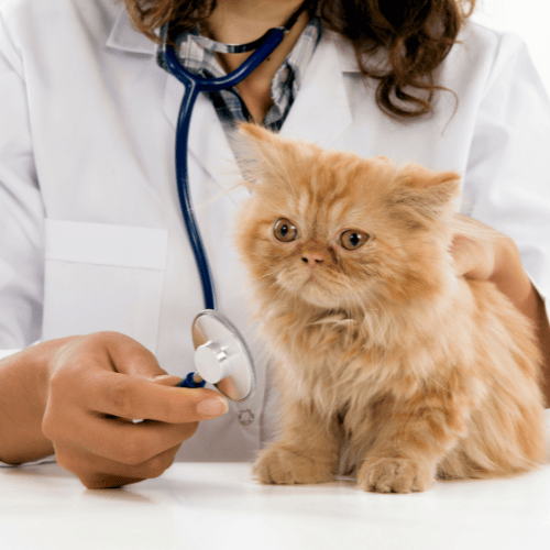
Pet Wellness
From new patient exams to pain management, we will provide the care your pet needs in our warm and inviting animal hospital.

Pet Medical
If your pet is acting lethargic or showing signs of illness, such as vomiting, diarrhea, or loss of appetite, we can get you the answers you need.

Pet Dentistry
Your pet’s oral health is an essential aspect of their overall wellness. Avoid gum disease and decay with dental cleanings for your dog or cat.
Emergency and After-Hours Care
For emergency care involving exotic pets (such as small mammals, birds, or reptiles), please call ahead to ensure that one of our exotic pet veterinarians are available. If not, our staff will direct you to the nearest exotic animal hospital.
Mesa Veterinary Hospital Team
Meet Our Veterinary Team

Katie Lawri, DVM
Dr. Lawrie was born outside of Toronto, Ontario. She attended Grand Valley State in Michigan on a swimming scholarship and completed a Bachelor of Science degree in Biology.
While applying to vet school she took a year off to work and spent about 3 months in South Africa working at West Coast National Park. She attended St George’s University in Grenada, West Indies for her doctor of veterinary medicine degree, ultimately completing that degree at Auburn University in Alabama.
She and her husband, who is also a veterinarian, moved to Colorado in 2023. Dr. Lawrie enjoys traveling, sewing, and spending time outdoors in her spare time.

Christine Horst, DVM
Veterinarian
Dr. Horst is from Golden, Colorado, and is a mom to two awesome boys. She tries to mountain bike as much as possible and she loves bike racing, skiing, and spending time with her family. Dr. Horst loves enjoying the outdoors with her kids. They currently share their home with two Bernese Mountain Dogs, a cat, and a guinea pig.
She attended Kent Denver School and upon graduation attended Colby College in Maine and graduated Phi Beta Kappa and Cum Laude. Dr. Horst then received her Doctor of Veterinary Medicine degree from CSU Veterinary School. She has been with the practice since 1999 and is a partner. Dr. Horst loves getting to know families and enjoys watching their pets grow old with them.

Kitren Nickerson, DVM
Veterinarian
Originally from Colorado Springs, Dr. Nickerson joined the Mesa Veterinary Hospital team in 2006. She is a practicing veterinarian and partner in the business. After earning her undergraduate degree from Cornell University and her Masters degree from Colorado State University, she went on to earn her Doctorate in Veterinary Medicine from Colorado State, graduating magna cum laude.
“I enjoy developing relationships with so many different people and their pets. I also love seeing new puppies and kittens that are starting life with their new families!”
Dr. Nickerson surrounds herself with animals at home, as well as at work. She has four dogs, three cats, and horses. She regularly competes in 3-day eventing of horses.
Shop Local
- Repeat deliveries
- Adjust any time
Thank you for supporting our practice in Golden, CO.
")